Atlas of Endoscopic Anatomy for Endonasal Intracranial Surgery
(Sprache: Englisch)

It is only recently that the use of the endoscope as the sole visualizing tool has been introduced in transsphenoidal pituitary surgery with its favorable related implications and minimal operative trauma. Of course, microscopic and endoscopic anatomy are...
Leider schon ausverkauft
versandkostenfrei
Buch (Kartoniert)
120.99 €
- Lastschrift, Kreditkarte, Paypal, Rechnung
- Kostenlose Rücksendung
- Ratenzahlung möglich
Produktdetails
Produktinformationen zu „Atlas of Endoscopic Anatomy for Endonasal Intracranial Surgery “
Klappentext zu „Atlas of Endoscopic Anatomy for Endonasal Intracranial Surgery “
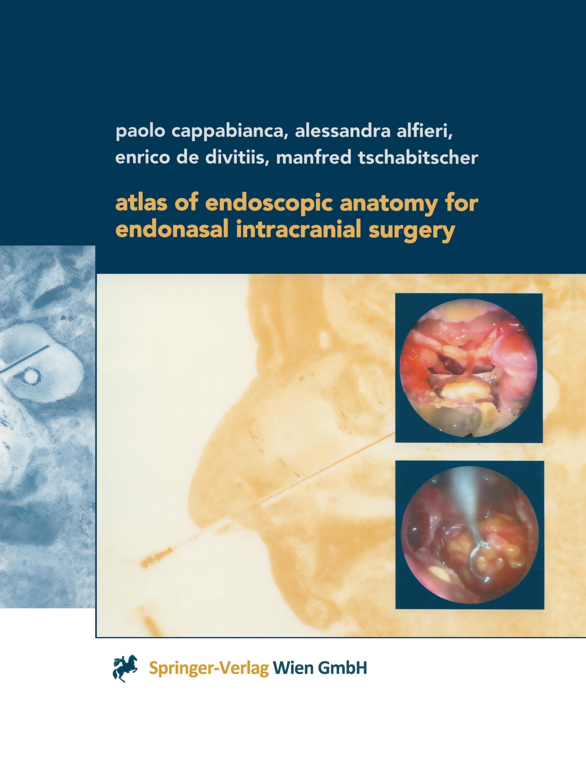
It is only recently that the use of the endoscope as the sole visualizing tool has been introduced in transsphenoidal pituitary surgery with its favorable related implications and minimal operative trauma. Of course, microscopic and endoscopic anatomy are basically the same, but the optical distorsion of endoscopic images is quite substantial compared to microscopic depictions. An endoscope lens produces images with maximal magnification at its center and severe contraction at its periphery. Nearer images are disproportionally enlarged and remote images are falsely miniaturized. This optical illusion may disorientate a surgeon who is not familiar with this peculiar condition at the skull base. This atlas acts as a guide through the endoscopic anatomy and gives detailed descriptions of the preoperative management and the surgical procedures.
Inhaltsverzeichnis zu „Atlas of Endoscopic Anatomy for Endonasal Intracranial Surgery “
I. Anatomic preparations.- I.A. Gross anatomy.- I.B. Endoscopic surgical anatomy.- II. Preoperative management.- II.A. Neuroradiological investigations.- II.B. Operating theatre.- III. Surgical procedure.- III.A. Surgical steps.- Appendix: Selected clinical cases.- Case 1: Intra-suprasellar macroadenoma.- Case 2: Intra-parasellar macroadenoma.- Case 3: Solid intra-suprasellar craniopharyngeoma.- Case 4: Cystic intra-suprasellar craniopharyngeoma.- Case 5: Arachnoid intra-suprasellar cyst.- Case 6: Intra-suprasellar RATHKE's cleft cyst.- References.
Bibliographische Angaben
- Autoren: Paolo Cappabianca , Alessandra Alfieri , Enrico de Divitiis , Manfred Tschabitscher
- 2012, Softcover reprint of the original 1st ed. 2001, XIII, 138 Seiten, Maße: 21 x 27,9 cm, Kartoniert (TB), Englisch
- Verlag: Springer
- ISBN-10: 3709172551
- ISBN-13: 9783709172551
- Erscheinungsdatum: 02.11.2012
Sprache:
Englisch
Rezension zu „Atlas of Endoscopic Anatomy for Endonasal Intracranial Surgery “
"... Den Autoren ist ein vom ausgezeichneten Bildmaterial dominierter Atlas gelungen ... " Annals of Anatomy 185/5/2003
Kommentar zu "Atlas of Endoscopic Anatomy for Endonasal Intracranial Surgery"
Schreiben Sie einen Kommentar zu "Atlas of Endoscopic Anatomy for Endonasal Intracranial Surgery".
Kommentar verfassen